The Manuel Lab focuses on unraveling the intricacies of the motor system and understanding how normal motor function is impacted in pathological conditions. Employing a range of cutting-edge techniques, including in vivo electrophysiology, in vitro electrophysiology, viral approaches, and advanced imaging methods, our research aims to shed light on the underlying mechanisms governing motor function and dysfunction.
Current Projects

Spinal Cord Physiology
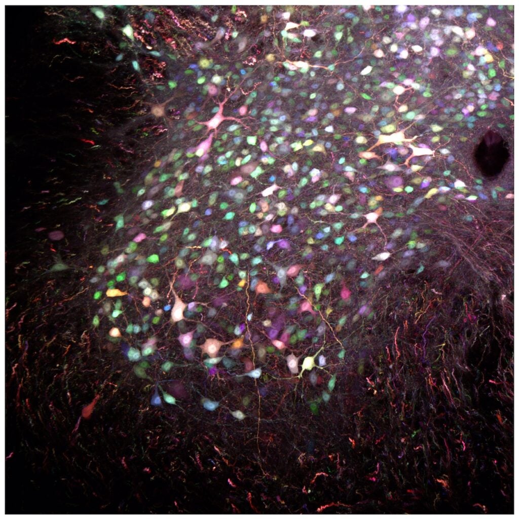
Our laboratory is dedicated to unraveling the intricate neural networks that orchestrate the production of movements. We investigate the finely tuned coordination and communication among motoneurons and interneurons within the spinal cord, aiming to decipher the underlying mechanisms that govern the generation and modulation of motor output. By delving into the complexities of these neural circuits, we seek to enhance our understanding of fundamental processes involved in movement production, laying the groundwork for potential interventions to address motor dysfunction in various pathological conditions.
Neurons in a section of mouse spinal cord, labeled using the brainbow technique. Image by Marin Manuel and Jean Livet; Institut de la vision, Paris, France

Amyotrophic Lateral Sclerosis (ALS)
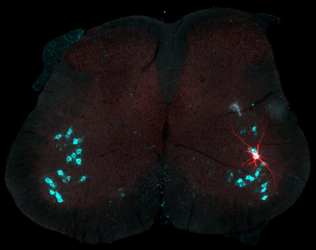
Our lab is dedicated to understanding how alterations in the electrophysiological properties of spinal motoneurons, and broader spinal networks, may contribute to the degeneration of motoneurons in the context of ALS. By exploring the intricate details of electrophysiological changes, we seek to uncover key insights into the mechanisms underlying ALS pathogenesis.
Spinal motoneuron recorded intracellularly and labeled by intracellular injection of Neurobiotin (red). Other motoneurons on this section of spinal cord labeled by immunolabeling against the choline-acetyl transferase enzyme (cyan). Picture by Marin Manuel and Rebecca Manuel.

Cerebral Palsy
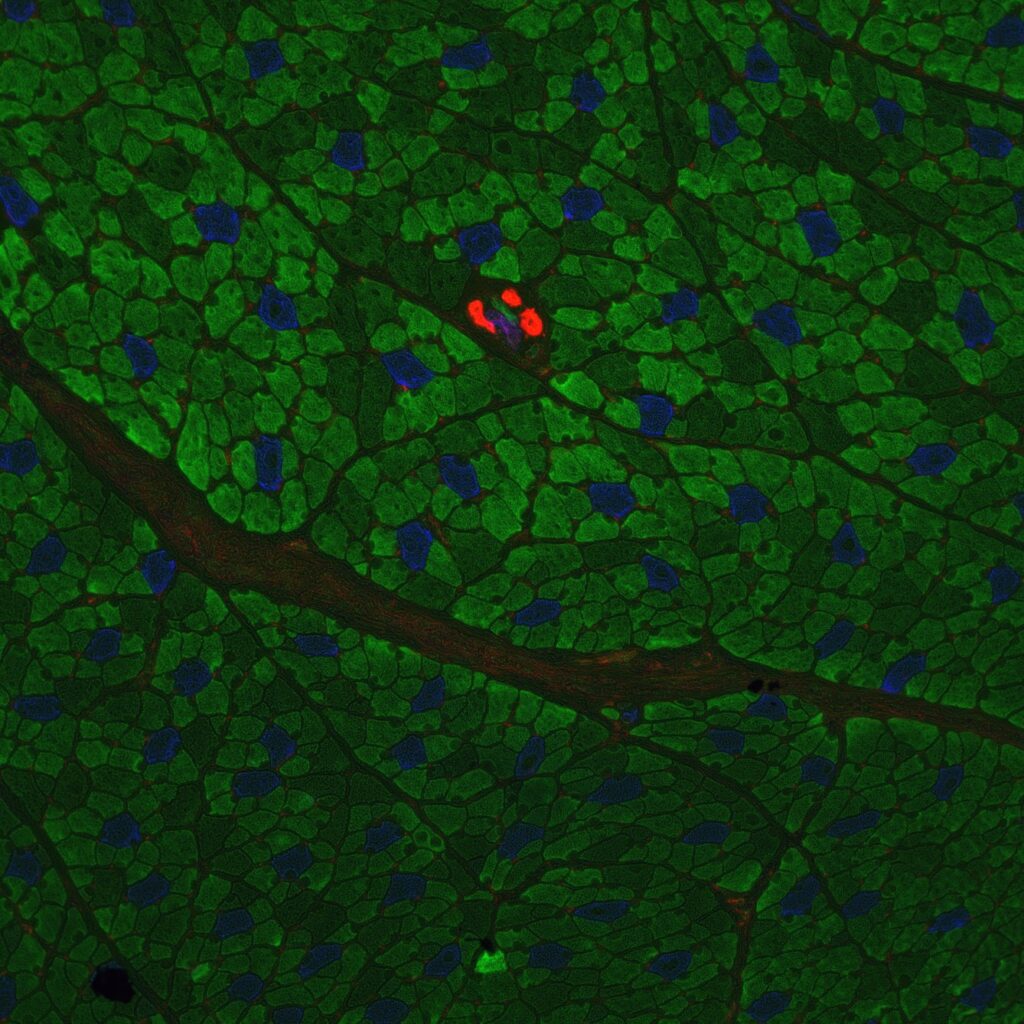
Focusing on the motor impairments associated with cerebral palsy, our research aims to uncover the specific electrophysiological alterations contributing to the condition. By utilizing advanced techniques, we strive to identify potential targets for intervention and therapeutic strategies to ameliorate motor dysfunction in individuals affected by cerebral palsy.
Cross section of a muscle from a rabbit model of CP. Image by Destiny Sanders and Emily Reedich.
The header image, “Fluorescence Microscopy of Neuromuscular Junctions,” was captured by Alyssa Madden ’23 and received an honorable mention in the URI Research and Scholarship Photo Contest.
